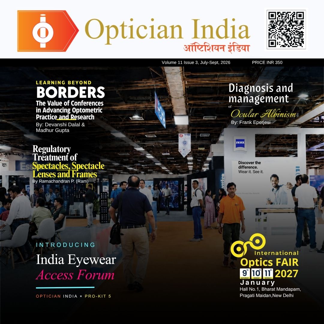
Diagnosis and management of ocular albinism

By: Dr. Frank Eperjesi
Ocular albinism is very rare but if it is missed at a young age it can have devasting consequences on education, activities of daily living and employment prospects.
There are two main types of albinism an optometrist may come across. Oculocutaneous albinism where the eyes, hair and skin are affected and ocular albinism where only the eyes are affected (iris, retina and choroid) and the hair and skin will have pigmentation often similar to that of the parents. A child with ocular albinism will not have the hall mark white hair and very pale skin associated with oculocutaneous albinism. This can make ocular albinism difficult to detect.
There is no medical treatment for ocular albinism but if it mistaken for bilateral amblyopia, because it causes high refractive error which may not be discovered until the child is three or four years-old (or sadly sometimes even later), the optometrist might think that glasses wear will help improve visual acuity. The optometrist might then recommend full-time glasses wear and review in three months without bringing in other experts to provide support. The visual acuity might improve a little over that three month period but as we see below there are anatomical reasons as to why it will not improve to levels that will allow age-expected progress in education and activities of daily living.
Cause of ocular albinism
It is caused by genetic mutations that disrupt the production and distribution of melanin pigment specifically in the eyes. The most common form of ocular albinism stems from mutations on the X chromosome. This impairs cellular structures called melanosomes, causing reduced pigmentation and poor eye development despite normal skin and hair pigmentation. The macula and fovea need normal pigment development and distribution in order to form a normal retinal structure. As it is an X-linked recessive mutation it is passed from mother to son and it almost exclusively affects males, while mothers carry the gene without showing any symptoms For a girl to have ocular albinism, she must inherit a changed gene on both of her X chromosomes. This requires a father who has ocular albinism and a mother who is at least a carrier, making cases in females very rare. The author was involved in a recent case of ocular albinism where the patient was a girl.
Symptoms of ocular albinism
Photosensitivity/photophobia and glare. As a reminder, photosensitivity/photophobia is a medical symptom defined as an abnormal intolerance or pain in the eyes and head caused by light, while glare is a visual sensation where bright light in the field of view reduces contrast or washes out visibility. In other words, photosensitivity/photophobia is a sensory-pain response, while glare is an optical or perceptual interference.
Clinical signs of ocular albinism
Below age-expected visual acuity.
Significant bilateral astigmatic hyperopia.
Pale iris/iris transillumination.
Manifest nystagmus possibly with a latent component.
Sometimes an abnormal head posture.
No or little retinal pigment epithelium and choroidal pigment.
No foveal reflex.
Central retinal vessels cross directly through the macula instead of forming a normal avascular zone.
Sometimes a strabismus.
Non-clinical signs
Not being able to see things/bumping into things.
Educational delay and difficulties with activities of daily living.
These symptoms and signs are addressed in more detail below.
Refractive error and visual acuity
Emmetropisation has failed and children with ocular albinism have bilateral high astigmatic hyperopia. In a recent case that the author was involved with, a five year-old girl had vision of 6/24 in each eye and a refractive error of right -3.00/+4.75 x 90 and left -1.50/+4.00 x 80. This was her first refraction and first pair of glasses. Her ocular albinism was not detected and she was not referred for low vision aids and educational support. After a year of full-time glasses wear and no other management her right visual acuity was 6/18 and left 6/15. Her visual acuity will never be better than this. Her ocular albinism was detected three-years later and she was referred to low vision and educational specialists.
Iris colour and iris transillumination
 |
The failure of ocular pigment production and distribution throughout the uvea (iris and choroid) leads to a pale iris such as that in the image above. In ocular albinism the skin and hair is normally pigmented and the iris is pale. Of course, there are many people with a pale iris who don’t have ocular albinism. It’s the presence of a pale iris and the symptoms and signs described in this article that assist in the diagnosis.
A common feature of ocular albinism is iris transillumination. This is caused by light going through the pupil, such as during retinoscopy or ophthalmoscopy, reflecting off the retina and passing out of the eye through those parts of the iris that have no or very little pigment. Examination of the eye using any light (even when minimal) will be very uncomfortable for a patient with ocular albinism.
 |
Example of iris transillumination
In everyday life, for a person with ocular albinism, light will go into the eye through the pupil and also through the iris where there are gaps in the pigment layer. This causes photosensitivity/photophobia and glare. Furthermore, as light impacts the retina it is not absorbed by the retinal pigment epithelium nor the choroid, because they are devoid of pigment so the light bounces around inside the eye off the white sclera and causes further problems with photosensitivity/photophobia and glare.
 |
Lack of retinal and choroidal pigment
Typical fundus appearance for a patient with ocular albinism. There is an obvious lack of retinal and choroidal pigment. The choroidal blood vessels and sclera are easily seen. Note, there is no foveal reflex and central retinal vessels cross directly through the macula instead of forming a normal avascular zone.
Macular hypoplasia and foveal hypoplasia
During foetal development the macula and fovea require pigment production and distribution in order to form properly. Irregular pigment formation and distribution prevent normal macular and foveal development. The macula and fovea become poorly differentiated, with the rods present more in the fovea and the cones distributed away from the fovea. The foveal pit may be reduced or not present (resulting in no foveal reflex), there is an absence of macular yellow pigment and the retinal vessels may cross through the macula. An under developed macula and fovea is termed macular and foveal hypoplasia. These are hallmark signs of ocular albinism.
Nystagmus
Manifest nystagmus possibly with a latent component will be present. This may be rotary or horizontal. It can have a small amplitude and high frequency making it difficult to detect. One good way is to look for subtle eye movements through a slit lamp using moderate to high magnification. Some people will also have a component of latent nystagmus where the amplitude and frequency increase when one eye is covered with an opaque occluder. This is important to recognise for the accurate measurement of visual acuity. If an opaque occluder is used to cover one eye in order to assess the visual acuity of the other eye and a person has latent nystagmus, on occlusion the amplitude of the nystagmoid movement will increase and the visual acuity will consequently reduce leading to an inaccurate measurement. The greater the nystagmoid movement the poorer the visual acuity. When a person has latent nystagmus, visual acuity can be measured by using a plus blurring lens over the eye that is not being measured instead of an opaque occluder. Using a plus blurring lens will not cause an increase in nystagmoid movement and accurate monocular visual acuity can be obtained.
Some children with manifest nystagmus due to ocular albinism may have a head-tilt, face-turn or chin-offset (abnormal head posture) as they have realised they can see better when adopting an abnormal head posture. Adopting an abnormal head posture dampens down or stops the nystagmoid movement and this abnormal head posture is called the null point. If a child is forced out of their abnormal head posture for cosmetic reasons the nystagmoid movement will increase in amplitude and frequency causing a reduction in visual acuity. When a person with nystagmus adopts an abnormal head posture it is better to let them adopt it.
Sometimes children with ocular albinism are inaccurately diagnosed as having congenital idiopathic nystagmus and not ocular albinism. This is probably because the nystagmus is detected and all the other signs typical of ocular albinism are missed.
Strabismus
In some cases of infantile nystagmus, individuals learn that if they overconverge they can see better because the overconvergence reduces the nystagmoid movement. While this ‘blockage’ successfully quiets the nystagmus to optimise visual acuity, it can result in a secondary esotropia. This clinical phenomenon is known as convergence dampening or nystagmus blockage syndrome. Because the eye oscillations slow down or stop during convergence, the eyes can focus on a target longer (increased foveation time). This significantly improves visual clarity. To maintain clearer vision a person may actively overconverge their eyes even when looking at distant objects. As well as constant overconvergence people will develop an abnormal head posture to compensate for the crossed position to align their preferred eye with the object of regard.
Management of ocular albinism
While the condition is permanent and there is no cure, it does not worsen over time. Parents/guardians can be educated about diagnosis, prognosis and recommendations.
In practice optical management focuses on improving visual acuity and ocular comfort and includes:
Prescription spectacles or contact lenses (when the patient can handle contact lenses and keep them clean) with prismatic correction in spectacle lenses in some cases of strabismus to maximise visual acuity.
Sunglasses and/or photochromic lenses to manage photosensitivity/photophobia and glare Consider specialised spectacles to assist with swimming and other sports.
If overconvergence causes vision problems, discomfort, or a significant abnormal head posture prescribing base-out prism that will cause the eyes to converge while looking straight ahead in order to dampen the nystagmus. Similar can be achieved through surgical intervention where extraocular muscle surgery can cause post-operative divergence. This structurally alters the resting muscle tension to naturally simulate the dampening effect of convergence in straight-ahead gaze.
When the child is mature enough to handle contact lenses and clean them, rigid gas permeable (RGP) contact lenses are a good option in terms of visual acuity and long term ocular health. Unlike spectacles, where the eyes move across and away from the optical centre, contact lenses sit directly on the cornea and move with every eye oscillation. This ensures the sharpest correction is always aligned with the visual axis. Because the image stays clear during movement, the brain has more stable moments (foveation periods) to process visual details. Contact lenses remove the peripheral prismatic effects, minification, magnification and blur caused by spectacle lenses.
The physical edge of a RGP lens gently interacts with the eyelids and conjunctiva during eye motion. This mild touch stimulates trigeminal nerve endings (afferent feedback pathways), which can reflexively help dampen the intensity and amplitude of the nystagmoid movement.
Rigid gas permeable lenses can also enhance low- and intermediate-frequency contrast sensitivity better than soft lenses or spectacles.
Types of management that require assistance from other experts are described below.
Low vision aids
The earliest possible referral when the child is of a young age allows for easier adaptation to low vision technology and enables the school system to serve the childs needs best, throughout their educational journey. Electronic low vision aids are portable (can be moved between classrooms) and can assist with near and distance tasks simultaneously.
 |
Example of a portable desk-top electronic low vision aid that can assist with distance and near tasks simultaneously.
 |
Example of a portable desk-top electronic low vision aid that can assist with near tasks.
 |
Hand held telescopes and stand and hand magnifiers can be used when away from the classroom, in the playground and on the way to and from school. A technology review every six months to make sure the child has the optimum devices should be advised.
Educational support
For children with low vision, educational support could include extended time on tests, use of technology and other low vision devices-as highlighted-an extra set of textbooks for home use, audio books, e-books, voice recognition software, a reading pen scanner and rest breaks during exams. Seating arrangements in the classroom, with a seat closer to the teacher and with fewer things around them and away from windows and bright lights can be discussed. Simpler and larger font size/style/colour in presentations and handouts, as well as reading tools that help children focus on specific lines will all help. Students with visual issues may benefit from increased hands-on learning and working in groups when instructions can be read out loud. Learning support every week with a focus on literacy and numeracy and learning to touch type are also useful.
Often there are national government agencies and/or NGOs that can provide low vision aids and educational support sometimes free of charge.
Sequence of management of ocular albinism
In practice optical management.
Advise parents/carers of the nature of the problem, the effect on visual acuity, photosensitivity/photophobia, glare, use of glasses full time, wear a peaked hat when in the sun, sit away from windows and other sources of light to reduce glare, referral pathways and possible future use of RGP contact lenses.
Provide information to teachers along with examples of the size of text the child can manage at distance and near with their glasses or contact lenses.
Referral to low vision specialist.
Referral to an educational specialist with experience of supporting children with low vision.
Diagnosis and management of ocular albinism
By: Dr. Frank Eperjesi Ocular albinism is very rare but if it is missed at a young age it can have devasting consequences on education, activities of da...
read more
Regulatory Treatment of Spectacles, Spectacle Lenses and Frames : Class A Exemption, Optical-Shop Dispensing and the Case for an Opticians Act
By P. Ramachandran (Ram) A Personal Industry Perspective Eyewear & Vision Care Industry Professional A discussion paper for consideration by India&...
read more
Beyond 20/20: Redefining Myopia Management and the Imperative Role of Indian Opticians
By: Vinay Swaroop Balla Navigating the 2050 Epidemiological Shift, Clinical Defocus Technologies, and the Paradigm Transition from Passive Dispensing t...
read more
37 years of specialty contact lens experience, brought to one of the most demanding ocular surface cases.
By GAGAN SAHNI Director – Professional Affairs Silverline Labs Pvt. Ltd Case Study Stevens-Johnson Syndrome : Mucous Membrane Grafting | Lid margin k...
read more
Industry Expert Spotlight: Subash Bansal Shares His Firsthand Experience with Nova MaxCvue Lenses
In the optical industry, few recommendations carry as much weight as those from seasoned professionals who experience products both as experts and daily wearers...
read more















.jpg)
.png)