From Classroom to Clinic: Learning Case of Keratoconus
.jpg)
By: Devanshi Dalal, Assistant Professor, Department of Optometry, BDIAS, CHARUSAT, Gujarat, Jaini Patel, Optometry Student, Department of Optometry, BDIAS, CHARUSAT, Gujarat.
Keratoconus is a progressive, non-inflammatory ectatic disorder of the cornea characterized by stromal thinning and conical protrusion, leading to irregular astigmatism, myopia, and visual distortion. It typically presents during puberty or early adulthood and may progress until the third or fourth decade of life. If undiagnosed or unmanaged in its early stages, it can severely impair quality of life due to significant visual disability.
Globally, the prevalence of keratoconus varies widely due to differences in diagnostic criteria and tools used. Earlier estimates suggested a prevalence of approximately 1 in 2,000 individuals, but newer studies utilizing corneal topography and tomography indicate that the condition may be far more common. In a landmark study conducted in South India using Scheimpflug imaging, Sridhar et al. (2022) reported a prevalence of 1 in 375 among school children, underscoring the need for early screening and diagnosis in high-risk populations.
The burden of keratoconus is particularly relevant in developing countries like India, where access to advanced diagnostic tools and specialty contact lens services may be limited. As optometry evolves, early detection and timely management of keratoconus have become key priorities, with optometrists playing a pivotal role in identification, patient education, contact lens fitting, and co-management with ophthalmologists.
Clinical Evaluation of Keratoconus: From Torchlight to Tomography
A thorough clinical evaluation of keratoconus (KC) represents a crucial component of optometric practice, especially as the profession moves from foundational learning in classrooms to hands-on clinical experience. Keratoconus, a progressive, non-inflammatory ectatic disorder of the cornea, often manifests subtly and can be mistaken for common refractive errors in its early stages. Early identification is vital to initiate timely interventions like corneal crosslinking and to prevent irreversible visual impairment. As clinical acumen develops, optometry students and practitioners learn to integrate a stepwise diagnostic approach—from basic chairside examination techniques like torchlight evaluation to advanced modalities such as corneal tomography. Each stage plays a unique and synergistic role in building a complete picture of the disease.
Torchlight Examination: Revealing the First Clues
In settings where high-end equipment is unavailable or during community eye care outreach, the simple torchlight becomes a vital screening tool. Despite its simplicity, it allows for the observation of subtle yet meaningful signs of corneal distortion. When viewed under oblique illumination, the cone-shaped protrusion of the cornea becomes more discernible, especially in moderate to advanced keratoconus. This forward bulging, although sometimes subtle, may cast an abnormal shadow—a phenomenon known as the oil-drop reflex, seen as a distorted red reflex through the pupil.
In advanced stages, Munson’s sign—characterized by a V-shaped indentation of the lower eyelid in downgaze due to the protruding cone—can be visualized using a basic torchlight. While these signs are not definitive, they serve as early indicators that merit further evaluation using more sophisticated instruments. Observational skills honed through basic examination methods prepare the clinician to suspect keratoconus even before objective data is available.
Retinoscopy and Refraction: Navigating the Optical Distortion
A key clinical challenge in early keratoconus is managing patients with frequent prescription changes or unexplained reductions in best-corrected visual acuity (BCVA). Retinoscopy remains an indispensable tool in this scenario, often revealing the classic scissoring reflex—a telltale sign of irregular astigmatism resulting from an uneven corneal surface. The reflex may appear distorted, flickering, or split, especially along oblique meridians.
Subjective refraction in keratoconus is notoriously challenging. Patients may exhibit large amounts of astigmatism—often oblique or against-the-rule—along with inconsistent endpoints. The presence of high-order aberrations further degrades the visual quality, leading to poor contrast sensitivity and fluctuating vision throughout the day. In the early stages, glasses may still offer functional correction; however, as the condition advances, conventional lenses fail to deliver acceptable clarity. This difficulty in achieving satisfactory correction should prompt a deeper corneal assessment.
Keratometry: Assessing Central Curvature
Manual and automated keratometers provide rapid, non-invasive assessment of central corneal curvature. While limited to approximately the central 3 mm of the cornea, these instruments still serve as valuable screening tools. Keratoconic corneas often yield steeper keratometric readings—exceeding 47.00 diopters in moderate cases and reaching above 60.00 diopters in severe stages. Additionally, keratometers may reveal irregular or fluctuating readings due to the distorted corneal surface, making consistent mire alignment difficult.
One of the limitations of keratometry is its inability to detect peripheral or inferiorly displaced cones, which are common in early keratoconus. Moreover, it offers limited data on the posterior corneal surface, a region increasingly recognized as critical in the earliest stages of ectasia. Thus, while keratometry serves as a useful tool in the diagnostic ladder, it necessitates follow-up with corneal topography or tomography for a comprehensive assessment.
Topography: Mapping the Irregular Landscape
Corneal topography has revolutionized the diagnosis and monitoring of keratoconus. Placido disc-based systems generate detailed curvature maps of the anterior corneal surface, enabling clinicians to detect localized steepening and asymmetry indicative of ectasia. The classic asymmetric bowtie or crab-claw patterns often indicate irregular astigmatism and thinning, particularly in the inferotemporal quadrant.
.jpg)
Quantitative indices such as the Inferior–Superior (I–S) value—a difference greater than 1.4 diopters between the inferior and superior curvatures—and the KISA% index (combining Keratometry, I-S, Skewed Radial Axis, and Astigmatism) have become diagnostic benchmarks. A KISA% value over 100% is strongly suggestive of keratoconus. These objective metrics aid in grading disease severity and identifying subclinical forms of the condition, especially in candidates for refractive surgery.
However, anterior surface maps only tell half the story. Posterior corneal elevation, which often precedes anterior changes, can go undetected without the use of tomographic systems such as the Pentacam or Galilei. These systems combine Scheimpflug imaging and slit scanning to provide a 3D model of the cornea.
Tomography and Pachymetry: Seeing Beneath the Surface
Tomographic imaging provides a more holistic view of the cornea, capturing both anterior and posterior elevation maps, as well as full-thickness pachymetric data. These devices allow clinicians to assess the shape, symmetry, and biomechanical behavior of the cornea with greater accuracy.
Pachymetry, the measurement of corneal thickness, plays a critical role in both diagnosis and management. In keratoconus, the cornea thins progressively, especially at the cone apex. Typically, the thinnest point is located inferotemporally and may be significantly displaced from the central axis. Accurate pachymetric data is crucial not only for confirming the presence of thinning but also for determining candidacy for interventions such as corneal collagen crosslinking (CXL), which requires a minimum corneal thickness threshold.
Modern Scheimpflug systems also generate indices like the Pachymetric Progression Index and Belin-Ambrosio Enhanced Ectasia Display (BAD-D), which are particularly useful in detecting forme fruste or subclinical keratoconus. Furthermore, the Tomographic Biomechanical Index (TBI) integrates shape and stiffness parameters to improve early detection sensitivity. These advancements bridge the gap between structural changes and biomechanical vulnerability, allowing for pre-emptive management decisions.
Optometrists serve as the first line of defense in the early identification and management of keratoconus. They play a vital role in screening high-risk individuals, such as adolescents with frequent prescription changes, and in educating patients about modifiable risk factors like chronic eye rubbing and allergic eye conditions. Their expertise in fitting specialty contact lenses—such as RGP, hybrid, and scleral lenses—provides functional vision to those with distorted corneas. Optometrists also co-manage advanced interventions like corneal crosslinking and post-surgical care, while monitoring disease progression through serial topography. Case-based learning further enhances our clinical judgment and nurtures essential soft skills like empathy and reassurance.
References
- Sridhar, S., et al. (2022). Prevalence and risk factors for keratoconus among school children in South India using Scheimpflug imaging. Indian Journal of Ophthalmology, 70(5), 1478–1483. https://doi.org/10.4103/ijo.IJO_1234_21
- Elsheikh, A., et al. (2023). AI-powered early detection of subclinical keratoconus using corneal imaging: A multicenter validation. Ophthalmology Science, 3(1), 100178. https://doi.org/10.1016/j.xops.2022.100178
- Hashemi, H., et al. (2021). Psychological impact of keratoconus and its management. Eye and Vision, 8(1), 19. https://doi.org/10.1186/s40662-021-00240-1
- Romero-Jiménez, M., et al. (2023). Genetic and molecular biomarkers in keratoconus: A systematic review. Experimental Eye Research, 227, 109350 https://doi.org/10.1016/j.exer.2023.109350
- Raiskup, F., et al. (2022). Accelerated versus standard crosslinking for keratoconus: A 2-year randomized study. Journal of Cataract and Refractive Surgery, 48(2), 159–166. https://doi.org/10.1097/j.jcrs.0000000000000789
2.jpg)
VISULUXE™ The Ultimate Progressive Lens Crafted for the Way Modern Life Moves
Introduction Life today demands constant visual transitions. A glance at a smartphone, a conversation across the room, reading messages, driving through traf...
read more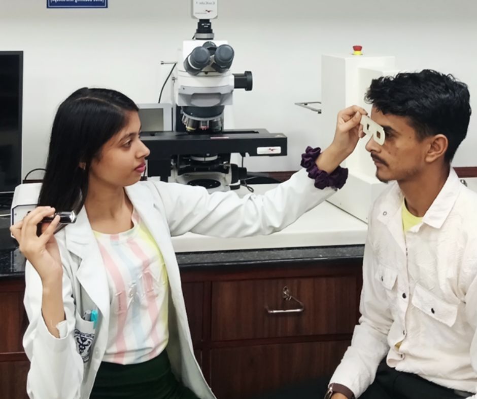
The Importance of Precise Pupillary Distance (PD) Measurement in Dispensing Eyewear
Article by: Sanjay K Mishra scientist Dr R P Centre AIIMS, New Dehli, Rajesh Kumar, Senior Optometrist, PGIMS, Univ of Health Sciences, Rohtak,&n...
read more3.jpg)
Uncorrected Refractive Error: India's Largest cost-effective Preventable Productivity Loss—and Its Greatest Growth Opportunity
By Ramachandran P. (Ram) India stands at a unique crossroads. With the world's largest population and one of its youngest workforces, the country has the...
read more1.jpg)
Cult glasses to have in your collection
Do you know the secret to always wearing the right glasses at the right time and with the right outfit ? No, it's not...
read more.jpg)
Untreated Cataract Can Lead to Blindness
Naushaba Afreen, B. Optometry (3rd year), Manipal Tata Medical College [MAHE] Introduction Cataract is the leading cause of preventable blindness worldwid...
read more.jpg)
.jpg)
.jpg)
.jpg)
.jpg)
.jpg)


1.jpg)


.jpg)
.jpg)


_(Instagram_Post).jpg)
.jpg)
_(1080_x_1080_px).jpg)


with_UP_Cabinet_Minister_Sh_Nand_Gopal_Gupta_at_OpticsFair_demonstrating_Refraction.jpg)
with_UP_Cabinet_Minister_Sh_Nand_Gopal_Gupta_at_OpticsFair_demonstrating_Refraction_(1).jpg)

.jpg)


.jpg)


.png)


