The Importance of Precise Pupillary Distance (PD) Measurement in Dispensing Eyewear
Article by: Sanjay K Mishra scientist Dr R P Centre AIIMS, New Dehli, Rajesh Kumar, Senior Optometrist, PGIMS, Univ of Health Sciences, Rohtak, Gauri Saini Optometry Student Dr R P Centre AIIMS, New Delhi Namrata Srivastav Associate Professor | HoD ERA University Lucknow, UP
As optometrists, we understand that dispensing a pair of glasses is a precise and scientific process. While the prescription (sphere, cylinder, and axis) is the primary determinant of visual acuity, the importance of accurate pupillary distance (PD) measurement cannot be overstated. PD, defined as the distance between the centers of the pupils,is a seemingly simple metric that profoundly impacts the comfort, visual performance, and overall satisfaction of the patient.This article delves into why precise PD measurement is essential and its impact on visual comfort and performance.
What is Pupillary Distance (PD)?
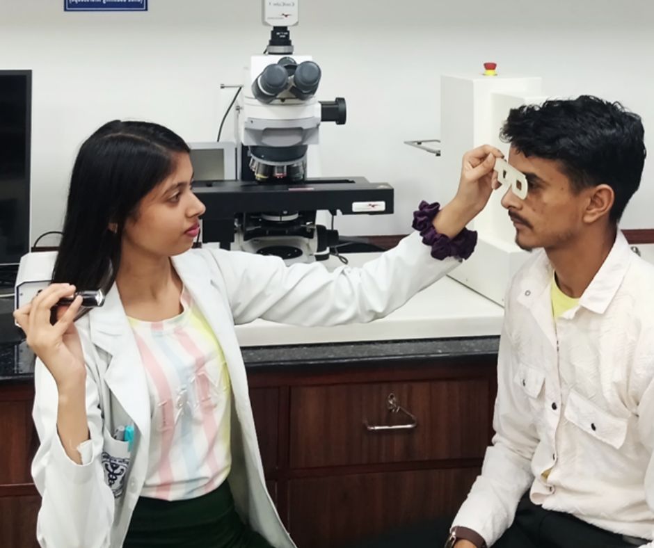
PD is the measurement of the distance, in millimeters (mm), between the centers of the pupils of the eyes. This measurement is crucial for correctly centering the optical center of the lenses over the patient’s pupils. When we look through a lens, the light rays are bent at the optical center, the point of no prism. If the optical center is not aligned with the pupil, a prismatic effect is induced, awhich can lead to various visual disturbances (figure 1).
The Impact of an Incorrect PD Measurement:
An inaccurate PD measurement can have several detrimental effects on the patient, ranging from mild discomfort to severe visual strain and headaches.
1. Induced Prism: The most significant consequence of an incorrect PD is the induction of unwanted prismatic effect. According to Prentice's Rule (figure 2), the amount of prism induced (P) is directly proportional to the power of the lens (F) and the decentration (c) in centimeters:
2025-09-10T11_11_17.jpg) |
For example, if a patient has a lens power of -5.00 D and the optical center is decentered by just 2 mm (0.2 cm), the induced prism is:
P=5.00×0.2=1.0Δ
This induced prism forces the extraocular muscles to work harder to fuse the two images, leading to asthenopia (eye strain). The higher the power of the prescription, the more critical the PD measurement becomes, as even a small decentration can cause a significant prismatic effect.
2. Visual Discomfort and Asthenopia: When the eyes are constantly fighting to overcome the induced prism, the patient experiences symptoms like eye fatigue, blurred vision, and headaches. This is particularly common in patients who wear their glasses for extended periods, such as those who work on computers or drive frequently. A patient with a seemingly perfect prescription may still complain of discomfort and headaches if the PD is off.
3. Compromised Visual Performance: Incorrectly centered lenses can degrade the quality of vision. The patient may not be looking through the optimal part of the lens, which can lead to peripheral distortion and reduced clarity. This can be especially noticeable in patients with high-index or aspheric lenses, where the optical design is highly sensitive to proper centration.
4. Binocular Vision Issues: An incorrect PD can disrupt the delicate balance of the binocular visual system. The induced prism can cause a vergence demand that the patient's fusional reserves may not be able to overcome, potentially leading to diplopia (double vision) or suppression of one eye. Over time, this can exacerbate or even induce latent phorias.
5. Patient Dissatisfaction and Redos: Ultimately, an inaccurate PD measurement can lead to patient dissatisfaction and the need for new glasses. This not only results in financial loss for the practice but also erodes patient trust. A redo due to a measurement error is entirely preventable and reflects poorly on the quality of care provided.
Methods for Accurate PD Measurement:
Given the critical nature of PD, optometrists must employ accurate and reliable measurement techniques.
1. Manual PD Ruler: This is the most traditional method. A PD ruler is held against the patient's brow, and the distance between the pupils is measured while the patient fixates on a distant object. While simple, this method is prone to human error, including parallax and inconsistent positioning.
2. Corneal Reflex Pupillometer: An improvement over the manual ruler, a pupillometer is an instrument that uses the patient’s corneal reflexes to precisely measure the distance between the pupils. This method is more accurate and less susceptible to parallax errors.
3. Digital and Automated PD Devices: Modern optical practices often utilize digital PD measurement devices or integrated systems within lensometers. These devices capturehigh-resolution images or videos of the patient's face and use algorithms to precisely calculate the PD. Many systems can also measure monocular PDs (the distance from the bridge of the nose to each pupil), which is essential for progressive and high-power prescriptions, as the nose is not always perfectly centered (figure 3).
2025-10-15T10_43_07.jpg) |
In the complex world of optometry, the importance of PD measurement should never be underestimated. It is a fundamental step in the dispensing process that directly impacts the patient visual comfort, performance, and overall satisfaction. A precise PD measurement ensures that the optical center of the lens is correctly aligned with the pupil, preventing the induction of unwanted prism and its associated symptoms. By utilizing accurate measurement techniques and understanding the implications of an incorrect PD, we can uphold our commitment to providing the highest standard of care to our patients, ensuring that their new glasses not only improve their vision but also enhance their quality of life.
.jpg)
True luxury? Time well spent and emotion
By Angelica Pagnelli In the past, "sister brands"—the segmentation of brands by major fashion houses—served to sell products to those w...
read more1.jpg)
Who Can Call Themselves an Optometrist? NCAHP May Finally Settle the Question
A personal perspective on professional standards, diploma qualifications and the future eye-care workforce By Ramachandran P. India's eye-care sector is...
read more.jpg)
Future Ready Optician: Trust, technology and the new language of luxury optical retail
By Moosi Kazmi True luxury in optics is personal accuracy. This is where the independent optician remains irreplaceable. A family returned to the s...
read more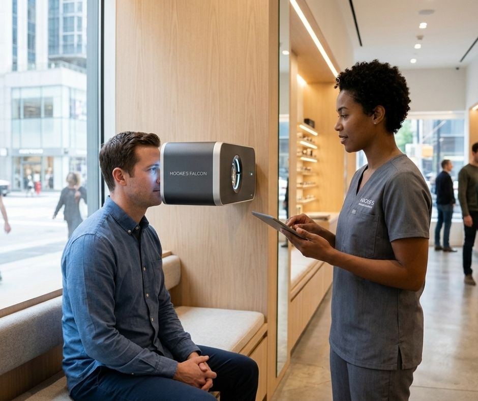
The Future of Eyewear in India: Premiumization, Technology, and Consumer Change
Interview with Akash Goyle In this exclusive interview, Madhur Gupta, Editor, Optician India sits down with industry pioneer Akash Goyle, the former Co...
read more1.jpg)
Childhood Myopia Enters Parliament: An Opportunity for India to Act
By Ramachandran P. Childhood myopia has finally received attention at one of the country’s highest policymaking forums. On 12 August 2026, Dr. Jas...
read more.jpg)
.jpg)
.jpg)
.jpg)
.jpg)
.jpg)

1.jpg)

.jpg)
.jpg)

_(Instagram_Post).jpg)
.jpg)
_(1080_x_1080_px).jpg)

with_UP_Cabinet_Minister_Sh_Nand_Gopal_Gupta_at_OpticsFair_demonstrating_Refraction.jpg)
with_UP_Cabinet_Minister_Sh_Nand_Gopal_Gupta_at_OpticsFair_demonstrating_Refraction_(1).jpg)
.jpg)




.jpg)

.png)