RAPD: Shining a Light on Hidden Ocular Conditions
_(4).jpg)
The pupillary light reflex is a critical component of the ocular examination and provides insight into the integrity of the afferent and efferent pathways of the visual system.
 |
A Relative Afferent Pupillary Defect (RAPD) or Afferent Pupillary Defect (APD) is a clinical sign indicating an asymmetric or unilateral defect in the afferent visual pathway, often affecting the optic nerve or retina. Identifying RAPD/APD is essential in diagnosing and managing various ophthalmic and neurological conditions.
TESTING FOR RELATIVE AFFERENT PUPILLARY DEFECT (RAPD)
RAPD occurs when one eye has reduced or absent afferent input relative to the other. The swinging flashlight test is the standard method for detecting RAPD.
Step-by-Step Procedure:
1. Preparation:
- Perform the test in a dimly lit room.
- Ensure the patient is seated comfortably and looking at a distant target to prevent accommodation.
- Use a bright, focused penlight.
2. Initial Observation:
- Observe the baseline size and symmetry of the pupils in ambient lighting.
- Check for anisocoria (unequal pupil sizes), which may indicate other conditions.
3. Direct and Consensual Reflex Testing:
- Shine the penlight directly into one eye for about 1 second.
- Observe the reaction of the illuminated eye (direct reflex) and the contralateral eye (consensual reflex).
- Repeat for the other eye.
4. Swinging Flashlight
 |
- Quickly alternate the penlight between the two eyes every 1–2 seconds.
- Observe the pupillary response as the light is moved from one eye to the other.
- Normal: Both pupils constrict equally when light is shone in either eye.
- RAPD: The affected eye’s pupil dilates (instead of constricting) when the light is moved to it, indicating reduced afferent input.
5. Grading the Severity of RAPD (optional):
- Use a neutral density filter to quantify the defect by placing filters of increasing density over the unaffected eye until the RAPD is no longer visible.
6. Record Findings:
- Document the presence and severity of RAPD, noting which eye is affected.
TESTING FOR AFFERENT PUPILLARY DEFECT (APD)
APD testing follows similar principles but may focus more on identifying structural lesions affecting the optic nerve or retina. Techniques like pupillography or infrared pupillometry can enhance sensitivity in APD detection.
 |
Key Steps:
1. Baseline Examination:
- Begin with the swinging flashlight test to identify asymmetry in the afferent pathway.
2. Alternative or Complementary Tests:
- Red Desaturation Test: Assess color vision by asking the patient to compare the intensity of red objects viewed through each eye.
- Visual Acuity and Visual Field Testing: Correlate APD findings with functional deficits.
3. Advanced Techniques:
- Use imaging (OCT or MRI) for structural evaluation.
- Conduct electrophysiological tests, such as Visual Evoked Potentials (VEP), for functional assessment.
CONDITIONS ASSOCIATED WITH RAPD/APD
- Optic Nerve Pathologies:
- Optic neuritis (commonly seen in multiple sclerosis)
 |
- Ischemic optic neuropathy
- Optic nerve compression (e.g., due to tumors)
- Retinal Pathologies:
- Severe retinal detachment
 |
- Central retinal artery or vein occlusion
- Neurological Conditions:
- Aneurysms or demyelinating diseases affecting the afferent pathway
CLINICAL SIGNIFICANCE
Testing for RAPD/APD is crucial in differentiating between optic nerve and ocular media diseases. A positive RAPD strongly suggests optic nerve dysfunction or severe retinal damage, even in cases where visual acuity appears normal. Early detection allows for prompt diagnosis and management of potentially sight-threatening or life-threatening conditions.
CONCLUSION
The swinging flashlight test remains the cornerstone for detecting RAPD, offering a quick, non-invasive method to assess the integrity of the afferent pathway. While advanced techniques and imaging modalities enhance diagnostic accuracy, a systematic approach to pupil testing can provide valuable insights into ocular and systemic health.
2.jpg)
VISULUXE™ The Ultimate Progressive Lens Crafted for the Way Modern Life Moves
Introduction Life today demands constant visual transitions. A glance at a smartphone, a conversation across the room, reading messages, driving through traf...
read more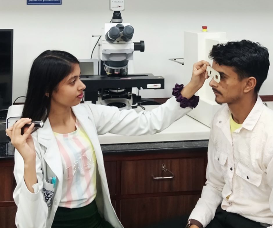
The Importance of Precise Pupillary Distance (PD) Measurement in Dispensing Eyewear
Article by: Sanjay K Mishra scientist Dr R P Centre AIIMS, New Dehli, Rajesh Kumar, Senior Optometrist, PGIMS, Univ of Health Sciences, Rohtak,&n...
read more3.jpg)
Uncorrected Refractive Error: India's Largest cost-effective Preventable Productivity Loss—and Its Greatest Growth Opportunity
By Ramachandran P. (Ram) India stands at a unique crossroads. With the world's largest population and one of its youngest workforces, the country has the...
read more1.jpg)
Cult glasses to have in your collection
Do you know the secret to always wearing the right glasses at the right time and with the right outfit ? No, it's not...
read more.jpg)
Untreated Cataract Can Lead to Blindness
Naushaba Afreen, B. Optometry (3rd year), Manipal Tata Medical College [MAHE] Introduction Cataract is the leading cause of preventable blindness worldwid...
read more.jpg)
.jpg)
.jpg)
.jpg)
.jpg)
.jpg)


1.jpg)


.jpg)
.jpg)


_(Instagram_Post).jpg)
.jpg)
_(1080_x_1080_px).jpg)


with_UP_Cabinet_Minister_Sh_Nand_Gopal_Gupta_at_OpticsFair_demonstrating_Refraction.jpg)
with_UP_Cabinet_Minister_Sh_Nand_Gopal_Gupta_at_OpticsFair_demonstrating_Refraction_(1).jpg)

.jpg)


.jpg)


.png)


