A-scan biometry and IOL master in axial length and intraocular lens power measurements at SITAPUR EYE HOSPITAL Uttarpradesh
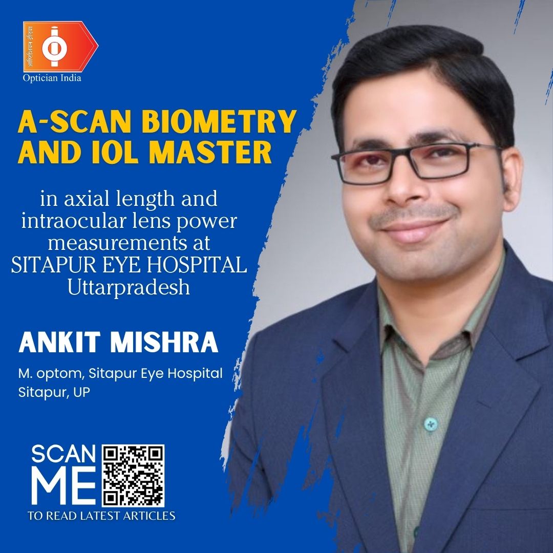
Purpose: The purpose of this paper is to evaluate differences between IOL master and A-scan regarding axial length (AXL) and predicted IOL power in different types of cataract.
Patients and methods: This study was conducted on 600 eyes of 600 patients (323 men and 227 women) during the period between 1st June 2017 to 20 Oct 2017 .The study patients were recruited from patients scheduled for phacoemulsification and IOL implantation.
The study patients who underwent routine ophthalmologic examination were informed about the purpose of the study and had to give an informed consent before inclusion.
Eyes with no ocular pathology apart from age-related cataract or complicated cataract to silicone injection or myopia were included in the study. The patients were divided into three groups including patients with SO-filled eyes who were scheduled for SO removal and phacoemulsification, high myopia with spherical equivalent (SE) greater than or equal to −6 D and or AXL greater than or equal to 26.0 mm, and eyes with nuclear cataract I and II.
The study excluded patients having retinal detachment, eyes whose AXL could not be measured with IOL master, because of dense ocular media opacities such as corneal scars or dense posterior sub-capsular cataracts, and patients who underwent previous corneal surgery (including refractive surgery). Patients having intraoperative complications such as inability to achieve secure ‘in the bag’ placement of the IOL (i.e. due to posterior capsule rupture, vitreous loss, weak zonules, or zonular rupture) were also excluded from the study.
Results: Six hundred eyes of 600 patients were included. The mean AXL measured by IOL master was higher (26.18±2.92 mm) than that with A-scan (25.02±2.99 mm) with a mean difference of 0.3±0.46mm (P=0.07). The mean predicted IOL power was 12.68±8.36 D with IOL master versus 12.09±8.23 D with A-scan (P=0.03). However, no statistically significant difference was found regarding average K-readings and predicted postoperative refraction (P=0.4 and 0.7, respectively). Bland–Altman plots showed almost perfect agreement between both methods regarding AXL and predicted IOL power. Further subgroup analysis revealed a statistically significant difference in AXL between both devices only in nuclear cataract with no significant difference in cases with complicated cataract to myopia or silicone oil (P=0.015, 0.3and 0.2, respectively). No statistically significant difference was found between the three groups regarding the calculated IOL power (P=0.36, 0.12 and 0.16, respectively). Bland–Altman analysis showed almost perfect agreement for the mean difference of AXL and IOL power in the three subgroups.
Conclusion: There is no significant difference between IOL master and A-scan biometry, with the non-contact IOL master being preferred by patients; however, there exists certain situations where A-scan is still mandatory.
Accurate calculation of the intraocular lens (IOL) power necessary for attaining the desired postoperative refraction is still a research issue which depends on several factors, including axial length (AXL), keratometry, anterior chamber depth and lens formulas. Of these factors, the preoperative measurement of AXL is considered to be a key determinant in calculating the IOL power to be implanted.
Studies based on preoperative and postoperative ultrasound biometry show that 54% of errors in predicted refraction after IOL implantation can be attributed to AXL measurement errors, 8% to corneal power measurement errors and 38% to incorrect estimation of postoperative anterior chamber depth.
Previously, applanation ultrasound (A-scan) biometry has been the most commonly used technique for AXL measurement. More recently, partial coherence laser interferometry has gained more preference.
The AXL when measured by applanation A-scan ultrasound causes erroneous AXL measurement and an undesired postoperative refractive outcome. This might be attributed to the indentation of the globe and an off-axis measurement of the AXL by the transducer particularly important in highly myopic eyes. IOL master is a fast, noncontact method reported as a potentially more accurate method than ultrasound biometry. IOL master uses the method of partial coherence interferometry (PCI) to measure the AXL, based on reflection of the interference signal of the retinal pigment epithelium. This technique was found to be more accurate than the acoustic method in cataractous eyes, with no other pathologies. It has also been suggested that the IOL master is more precise and useful in difficult situations, including high myopia, posterior staphyloma or silicone oil (SO)-filled globes.
The AXL measurement with the IOL master is not affected by the subjective error sources of acoustical A-scan ultrasound biometry. Measurement along the visual axis is ensured as the patient fixates on the light source, precluding a misalignment error produced by an off-axis posterior staphyloma. However, it will not work in the presence of significant axial opacities. A mature or darkly brunescent lens, dense posterior subcapsular plaque, vitreous haemorrhage or central corneal scar will preclude any type of meaningful measurement. On the other hand, eyes with posterior staphyloma, or eyes with SO, are very easy, and routinely measured with the IOL master.
 |
The aim of this work was to determine whether IOL power calculations for cataract surgery as measured by postoperative refractive error using PCI are more accurate in improving postoperative outcomes than applanation ultrasound biometry in high myopic patients, SO-filled eyes, and in nuclear cataracts (I and II).

.jpg)
.jpg)
.jpg)
.jpg)


1.jpg)



.jpg)
.jpg)


_(Instagram_Post).jpg)
.jpg)
_(1080_x_1080_px).jpg)


with_UP_Cabinet_Minister_Sh_Nand_Gopal_Gupta_at_OpticsFair_demonstrating_Refraction.jpg)
with_UP_Cabinet_Minister_Sh_Nand_Gopal_Gupta_at_OpticsFair_demonstrating_Refraction_(1).jpg)
.jpg)






.jpg)



.png)
