Effectiveness of Auto refractor as a first level screening method or irregular astigmatism compared to the Photokeratoscopy - A cross sectional study
ABSTRACT
AIM: To present a method of screening for irregular astigmatism with an auto refractor and its determinants compared to photokeratoscopy.
METHODS: This cross-sectional validity study was conducted at Sitapur eye hospital. A tabletop auto refractor (Test 1) was used to measure the refractive status of the anterior surface of the cornea at two corneal meridians of each eye. Then photokeratoscopy (Test 2) and Bogan’s classification was used to group eyes into those with regular or no astigmatism (GRI) and irregular astigmatism (GRII). Test 1 provided a single absolute value for the greatest cylinder difference (Vr). The receiver operating characteristic (ROC) were plotted for the Vr values measured by Test 1 for GRI and GRII eyes. On this basis a Vr value was cut off and sensitivity & specificity was also calculated.
RESULTS: The study sample was comprised of 260 eyes (135 patients). The prevalence of irregular astigmatism was 42% [95% confidence interval (CI): 36, 48]. Based on] Test 2, there were 151 eyes in GRI and 109 eyes in GRII. The median Vr was 0.75 D (25% quartile, 0.5 D) for GRI and 1.75 D (25% quartile, 1.25 D) for GRII. The area under curve was 0.171 for GRI and 0.83 for GRII. The sensitivity of test I was 78.1% and the specificity was 76.1%.
CONCLUSION: A conventional auto refractor can be effective as a first level screening method to detect irregular corneal astigmatism in places where photokeratoscopy facilities are not available.
KEYWORDS: screening; irregular astigmatism; auto refractor; photokeratoscopy; cornea; validity.
INTRODUCTION:
The volume of refractive surgery has steadily increased over time. Hence, detection and management of irregular astigmatism has become crucial for improving outcomes and for patient satisfaction. However, as the volume of refractive surgery increased over the last 2-3 decades, a diagnosis of irregular astigmatism has become more common[1- 2]. Additionally, better detection of irregular corneas has become paramount for modern cataract surgery. Prior to the introduction of photokeratoscopy, irregular astigmatism was diagnosed with scissors movement on retinoscopy and/or deformation of the mires during manual keratometry [2]. However, a keratometer only provides a crude, qualitative measure of irregular astigmatism, subjectively judged by distortion of the mires[3]. Although keratometry provides information on corneal image forming properties, such as corneal astigmatism, it is inaccurate for irregular astigmatism. Irregular astigmatism can also be suspected in cases with impaired vision that is corrected by placement of rigid contact lenses [2]. However, rigid contact lens fitting causes patient discomfort and involves significant patient chair time precluding its use as a diagnostic test for irregular astigmatism. Proper placement of a pinhole to align with the visual axis can yield accurate visual acuity. However, this is a subjective test that precludes diagnosis of peripheral irregularities and is influenced by other conditions such as retinal damage and cataracts [4].
Photokeratoscopy provides the most comprehensive information on corneal regularity and curvature for the diagnosis of irregular astigmatism [3,5-6]. However, availability, cost and the ability to interpret the outcomes is a challenge in all ophthalmic clinics. Tabletop auto refractors are available in most ophthalmic and optometric clinics. Auto refractors acquire measurements rapidly and are patient friendly to use compared to topography [7-8]. The aim of this study is to validate a quantitative test for irregular astigmatism using an autorefractor compared to conventional photokeratoscopy.
SUBJECTS AND METHODS:
This was a non-randomized, cross sectional study of consecutive patients who presented to a public general ophthalmology outpatient clinic in Sitapur eye Hospital, from June to August 2021. Patients were included after informed consent was obtained. Patients were excluded if they had mental or physical disability, uncooperative, history of recent ocular surgery, corneal scars or acute ocular pathology at examination.
Patients’ data were collected on demographics such as age, gender and laterality of the condition and the distance visual acuity with and without spectacles. Distance vision was tested monocularly using a Snellen illiterate C visual acuity chart held at 6m distance. An optometrist performed dynamic refraction for each eye without pharmacologic cycloplegia.
Methods to Detect Irregular Astigmatism
Test 1: Astigmatism evaluation using an objective asymmetric refractometer was performed with a tabletop autorefractor (Topcon KR 800, Topcon Corp.) to determine the refractive status of anterior surface of the cornea in two meridians that pass through the pupil. P and P' are symmetrical points located at opposite ends of a symmetric corneal parallel. Stated differently, P and P' are diametric opposite ends of an ellipse, hence, both have the same curvature as the parallel. Consequently, if two symmetrical points of a cornea have the same radius as the meridian and also the same radius as the parallel, its astigmatism will be regular.
Four measurements were performed within the pupillary area near the iris sphincter at 45°, 135°, 225° and 315° meridians (Figure 1). All measurements were obtained by an experienced operator using the same machine and procedure. Subjects were instructed to look at an optically distant target displayed in the autorefractor and keep their eyes wide open during this measurement. All refractions were noted in negative cylinder notation. The readings were recorded, 1-2s after a blink. Average values of the refraction measurements were noted from the auto refractor and were recorded using an absolute magnitude of cylinder notation.
At the 45° meridian, we performed measurements number 1 and 3 (45º and 225º). We termed this V45 which was the absolute value of the difference between the first measurement of astigmatism (Cyl_145) and the third (Cyl_3225) so that V45=Cyl_145 -Cyl_3225.
At the 135º meridian, we performed measurements number 2 and 4 (135º and 315°). This was termed V135, the absolute value of the difference between the astigmatism of the second measurement (Cyl_2135) and the fourth (Cyl_4315) so that V135=Cyl_2135 -Cyl_4315.
We choose the highest absolute value between V45 & V 135 and termed it Vr. Vr designated the greater asymmetry in the cornea. To obtain a cut-off value, all the Vr values were compared to our topographic classification.
 |
Figure 1 Schematic drawing of Test 1
Method A: Each meridian of a cornea formed of two semi-meridians symmetrical from the optical axis, any two points Pand P’ of these semi- meridians located at the same distance from the corneal apex has the same curvature;
Method B: Four measurements were performed within the pupillary area near the iris sphincter at 45°, 135°, 225° and 315° meridians.
Example for right eye (OD):
Measurements with refractometer in OD; measure at 45º=-1, -1×45º, measure at 225º=-0.5, -1×225º, measure at 135º=1, -0.5×135º, measure at 315º=-0.5, - 2×315º; Then we have the absolute value of the cylinder value of each meridian, getting Cyl45=1, Cyl225=1, Cyl135=0.5, Cyl315=2; Calculate V45=0, V135=-1.5; Calculate Vr we choose the highest absolute value between V45 and V135.
The eyes were separated into two groups, based on the photokeratoscopy classification: group I [regular or no astigmatism (GRI)] including eyes with normal or regular astigmatism (round or oval and symmetric pattern); and group II [irregular astigmatism (GRII)], containing eyes with irregular astigmatism (irregular and unclassified) considering asymmetric photokeratoscopy images 9. Eyes were qualitatively classified based on Bogan’s recommendations [9].
To minimize variation in the results, all measurements were performed between 9 a.m. and 2 p.m. The examiner and participants were masked to the results of the previous measurements obtained from each device. Participants were instructed to blink completely just before each measurement. They were asked to sit back after each repeat measurement, and the device was realigned before each measurement.
The effectiveness of auto refractors in defining irregular astigmatism in both GRI and GRII was compared to that found by photokeratoscopy was performed using the receiver operator characteristic (ROC) curve. The results of this analysis was used to determine the diagnostic cut-off points (Vr=1.25 D) to determine the overall predictive accuracy of the test as described by the area under the curve (AUC). These curves are obtained by plotting sensitivity against 1-specificity, calculated for each value observed. An area of 100% implies that the test perfectly discriminates between groups. We also used this approach to calculate specificity, sensitivity. The validity parameters were sensitivity, specificity, positive predictive value, negative predictive value and prevalence of irregular astigmatism. The 95% confidence interval (CI) of the validity parameters was also calculated. P<0.05 was considered statistically significant.
RESULTS
_(1)001.jpg) |
_(1)000.jpg) |
The study sample consisted of 260 eyes of 135 participants. There were 58 (43%) males and 77 (57%) females. The median age of participants was 35.5y (25% quartile, 25y). There were 132 (50.8%) right eyes and 128 (49.2%) left eyes.
Based on the Bogan et al [9] corneal photokeratoscopy classification, 151 (58%) eyes had no astigmatism or regular astigmatism (GRI) and 109 (42%) eyes had irregular astigmatism (GRII). The Comparison of auto-refractor measurements in eyes with and without irregular astigmatism is presented in Table 1. The Vr values were significantly higher in GRII compared to GRI. The ROC of GRI and GRII is presented in Figure 2. The AUC in GRI and GRII were 0.17 and 0.83 respectively.
The validity parameters for Test 1 were estimated by comparing the presence and absence of irregular astigmatism as defined by Test 2. Irregular astigmatism was defined as a Vr value greater than 1.25 D. The sensitivity, specificity were calculated using standard formulas (Table 2).
_(2).jpg) |
Table 2: Validity of Astigmatism screening by Auto refractor compared to photokeratoscopy Sensitivity: 118/151×100%=78.1% (95% CI 73.1, 83.1); Specificity: 83/109×100%=76.1% (95% CI 71.0, 81.3); False p o s i t i v e s : 26/144×100%=18.1% (95% CI 13.4, 22.7); False negatives: 33/116×100%=28.4% (95% CI 23.0, 33.9).Prevalence of Irregular astigmatism: 109/260×100%= 42%.
|
_(3).jpg) |
Figure 2: Area of ROC curve (graphical plot of the sensitivity vs. 1-specificity) for astigmatism the cut-off was Vr 1.25 D, with 78.1% sensitivity and 76.1%specificity.
We also studied the influence of age-group, gender and the eye involved on the validity parameters such as sensitivity and specificity of auto-refractor screening (test 1) for irregular astigmatism (Table 3). Age group was statistically significantly positively associated with specificity (P<0.001) and negatively associated with sensitivity (P=0.006). However female gender (P=0.008) and left eyes (P=0.05) had statistically significantly higher specificities compared to males and right eyes.
_(4).jpg) |
Table 3: Variation in validity parameters of greatest cylinder value in two diagonal meridians by an autorefractor compared to photokeratoscopy by determinants.
DISCUSSION
This study is unique as it attempted to evaluate the utility and reliability of an autorefractor, a commonly available diagnostic tool for basic refractive examination, as a method for screening irregular astigmatism. Auto refractors are inexpensive and routinely used in most clinics. The purpose of our investigation was to show that it can be use in triage to identify patients who require photokeratoscopy to confirm the diagnosis of irregular astigmatism and further management. Astigmatism is a clinically important condition and accounts for about 13% of the refractive errors of the human eye [10].
Astigmatism greater than 1D cylinder represents significant irregularity 10-11-12 In our participants 42% had irregular astigmatism but the current study was done using a not randomized population sample and data were based on a convenience sample, composed of individuals who spontaneously requested ophthalmic treatment. Hence the prevalence of astigmatism reported in this study must be interpreted with caution. The high prevalence could be because our ophthalmic clinic is known in the region for its expertise in dealing with keratoconus patients and referral bias may play a role.
In our study there was no association between irregular astigmatism and gender. This result need to be seen with caution since our sample had enrolled more females. However, it remains unclear if gender is determinant in the astigmatism prevalence [13-14] or the preponderance of keratoconus [15-16].
Mainly young patients (below 20y) had high corneal astigmatism that decreased with age [12]. We elected to study Vr, based on each meridian formed by two semi-meridians symmetrical to the optical axis. These are two points, P and P' of these semi-meridians located at the same distance from the corneal apex and have the same curvature. In a normal cornea all meridians are elliptical curves and the curvature of the meridian varies in a mathematically predictable manner as the distance from the corneal center increases [17].
For irregular astigmatism, it is highly unlikely that the radii of curvature of the meridian and the parallel of these two symmetrical points are equal, so we can assume the pairs of symmetric points of a cornea with irregular astigmatism do not have the same astigmatism. As expected, the Vr value in our sample was significantly higher in GRII, the group with irregular astigmatism. GRII had significant differences in cylinders in different points of the cornea, designated by the greater asymmetry in the cornea.
The overall predictive accuracy of Vr, as described by the area under the ROC curve (AROC), was high in GRII (0.83) with values >0.9[29]. Hence,test1 withVr was effective for the screening for irregular astigmatism. The cut-off point of Vr 1.25 D showed high sensitivity and specificity (78.1% and 76.1%, respectively).
In contrast, our method is an objective quantitative method, which is quick, easy and reliable for screening for irregular astigmatism. We obtained a Vr value which designated the greater asymmetry in the cornea, by only using an autorefractor. This new proposed method is adequate for primary screening but has some limitations. To perform test 1 correctly, the autorefractor should be directed exactly on 2 pairs of symmetrical points on the cornea in the same meridian. However, we recognize that in practice, perfect symmetry is difficult to achieve. Therefore, we accept that the points can be similar distances from the centre of the cornea in the same meridian. As a consequence of the difficulty in measuring at perfectly symmetric points in the same meridian, applying test 1 to corneas with irregular astigmatism, the possible error will be added or subtracted to the actual differences that may exist. However, this error also exists in Photokeratoscopy, as it is impossible to take two photokeratoscopy that are precisely aligned.
In conclusion, test 1 is not designed to compete with the Photo-keratoscopy. It offers the possibility of a likely diagnosis applicable to all patients presenting to a general ophthalmology/ Optometrist clinic. Our method permits the identification of cases suspicious for irregular astigmatism and those should undergo photokeratoscopy. Hence this is a screening tool for patients who require further workup. This optimizes the use of the photokeratoscopy and allows for greater clinical efficiency. Although it cannot be concluded from this study that Vr is sufficient alone as a single diagnostic index, it does seem to be very effective in discriminating irregular from regular astigmatism. Thus data concerning Vr >1.25 D should be combined with curvature data in stratifying patients with this condition.
REFERENCES
1. Nordan LT. Keratoconus: diagnosis and treatment. Int Ophthalmol Clin 1997;37(1):51-63.
2. Goggin M, Alpins N, Schmid LM. Management of irregular astigmatism. Curr Opin Ophthalmol 2000;11(4):260-266.
3. Roh HC, Chuck RS, Lee JK, Park CY. The effect of corneal irregularity on astigmatism measurement by automated versus ray tracing keratometry. Medicine (Baltimore) 2015;94(13):e677.
4. Mohammad-Rabei H, Shojaei A, Aslani M. Concurrent macular corneal dystrophy and keratoconus. Middle East Afr J Ophthalmol 2012;19(2): 251-253. 5 McMahon TT, Anderson RJ, Joslin CE, Rosas GA. Precision of three topography instruments in keratoconus subjects. Optom Vis Sci 2001; 78(8):599- 604
5. Stefano VS, Melo Junior LA, Mallmann F, Schor P. Interchangeability between Placido disc and Scheimpflug system: quantitative and qualitative analysis.
6. Arq Bras Oftalmol 2010;73(4):363-366. Lowry EA, de Alba Campomanes AG. Cost-effectiveness of school- based eye examinations in preschoolers referred for follow-up from visual screening.
7. JAMA Ophthalmol 2016;134(6):658-664 Williams C, Lumb R, Harvey I, Sparrow JM. Screening for refractive errors with the Topcon PR2000 pediatric refractometer. Invest Ophthalmol Vis Sci2000;41(5):1031-1037
8. Bogan SJ, Waring GO 3rd, Ibrahim O, Drews C, Curtis L. Classification of normal corneal topography based on computer-assisted video-keratography. Arch Ophthalmol 1990;108(7):945-949.
9. Porter J, Guirao A, Cox IG,
10. Williams DR. Monochromatic aberrations of the human eye in a large population. J Opt Soc Am A Opt Image Sci Vis 2001;18(8):1793-1803.
11. Parssinen O. Astigmatism and school myopia. Acta Ophthalmol (Copenh) 1991;69(6):786-790.
12. Marasini S. Pattern of astigmatism in a clinical setting in Maldives. J Optom 2016;9(1):47-53
13. Gordon-Shaag A, Millodot M, Shneor E, Liu Y. The genetic and environmental factors for keratoconus. Biomed Res Int 2015;2015: 795738.
14. Bawazeer AM, Hodge WG, Lorimer B. Atopy and keratoconus: a multivariate analysis. Br J Ophthalmol 2000;84(8):834-836.
15. Saini JS, Saroha V, Singh P, Sukhija JS, Jain AK. Keratoconus in Asian eyes at a tertiary eye care facility. Clin Exp Optom 2004;87(2):97-101.
16. Fink BA, Wagner H, Steger-May K, Rosenstiel C, Roediger T, McMahon TT, Gordon MO, Zadnik K. Differences in keratoconus as a function of gender. Am J Ophthalmol 2005;140(3):459-468
17. Lindsay R, Smith G, Atchison D. Descriptors of corneal shape. Optom Vis Sci 1998;75(2):156-158
2.jpg)
VISULUXE™ The Ultimate Progressive Lens Crafted for the Way Modern Life Moves
Introduction Life today demands constant visual transitions. A glance at a smartphone, a conversation across the room, reading messages, driving through traf...
read more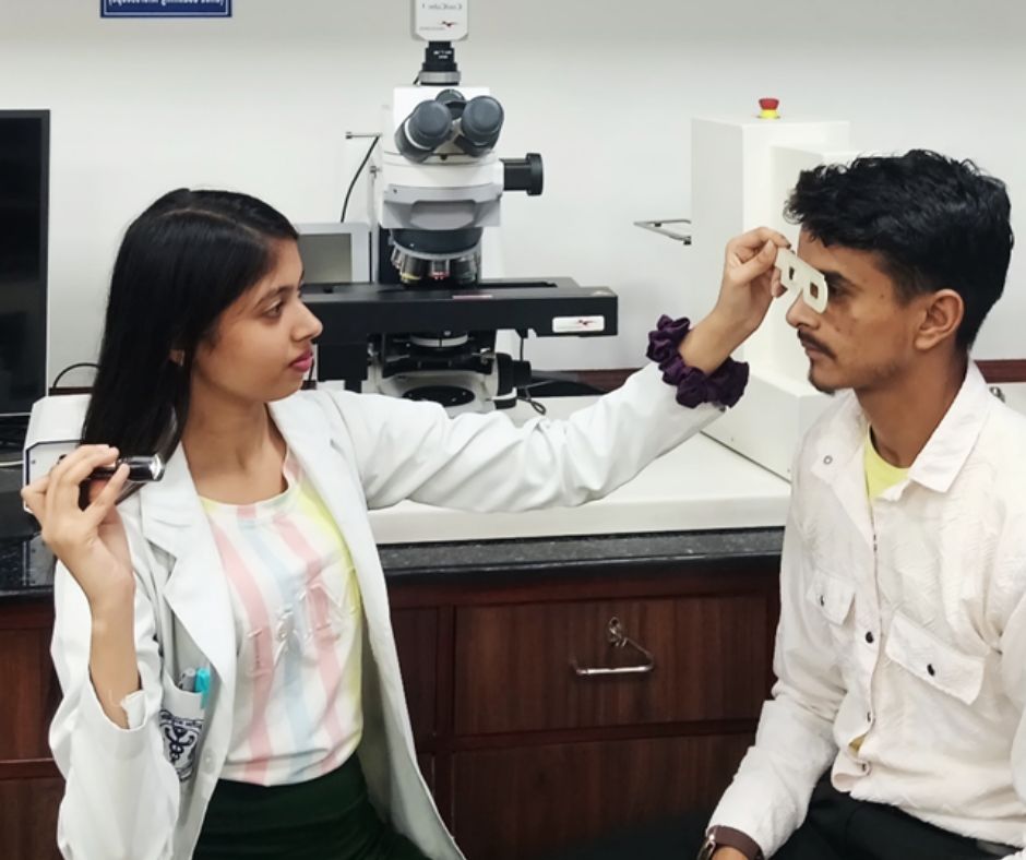
The Importance of Precise Pupillary Distance (PD) Measurement in Dispensing Eyewear
Article by: Sanjay K Mishra scientist Dr R P Centre AIIMS, New Dehli, Rajesh Kumar, Senior Optometrist, PGIMS, Univ of Health Sciences, Rohtak,&n...
read more3.jpg)
Uncorrected Refractive Error: India's Largest cost-effective Preventable Productivity Loss—and Its Greatest Growth Opportunity
By Ramachandran P. (Ram) India stands at a unique crossroads. With the world's largest population and one of its youngest workforces, the country has the...
read more1.jpg)
Cult glasses to have in your collection
Do you know the secret to always wearing the right glasses at the right time and with the right outfit ? No, it's not...
read more.jpg)
Untreated Cataract Can Lead to Blindness
Naushaba Afreen, B. Optometry (3rd year), Manipal Tata Medical College [MAHE] Introduction Cataract is the leading cause of preventable blindness worldwid...
read more.jpg)
.jpg)
.jpg)
.jpg)
.jpg)
.jpg)


1.jpg)


.jpg)
.jpg)


_(Instagram_Post).jpg)
.jpg)
_(1080_x_1080_px).jpg)


with_UP_Cabinet_Minister_Sh_Nand_Gopal_Gupta_at_OpticsFair_demonstrating_Refraction.jpg)
with_UP_Cabinet_Minister_Sh_Nand_Gopal_Gupta_at_OpticsFair_demonstrating_Refraction_(1).jpg)

.jpg)


.jpg)


.png)


